Ultrasound-Guided IV Insertion Training for Nurses: A Comprehensive Plan
This plan details a structured curriculum for nurses‚ utilizing simulation with silicone and tofu models‚ to achieve mastery in ultrasound-guided intravenous (USGIV) access.
Ultrasound-Guided Intravenous (USGIV) insertion represents a paradigm shift in peripheral intravenous (PIV) access‚ moving beyond palpation-based techniques. This method employs real-time ultrasound visualization to identify suitable veins‚ enhancing insertion success and minimizing complications. Traditionally‚ nurses relied on tactile feel and visual assessment‚ often leading to multiple attempts and patient discomfort.
USGIV training equips nurses with the skills to confidently locate and cannulate veins‚ even in challenging patients – those with obesity‚ edema‚ or difficult anatomy. The adoption of USGIV is driven by a desire to improve patient safety‚ reduce healthcare costs associated with failed attempts‚ and elevate the standard of IV therapy. This comprehensive plan aims to provide a robust educational framework for nurses to integrate USGIV into their practice.
II. The Need for USGIV Training in Nursing
The increasing complexity of patient care and a growing emphasis on patient safety necessitate widespread USGIV training for nurses. Traditional IV insertion often results in significant rates of failure‚ causing patient discomfort‚ delayed medication administration‚ and increased healthcare costs. Multiple attempts heighten the risk of phlebitis‚ infiltration‚ and nerve damage.
Furthermore‚ a significant portion of the patient population presents with anatomical challenges – obesity‚ edema‚ or compromised venous access – making traditional methods unreliable. USGIV offers a solution by providing real-time visualization‚ allowing nurses to confidently locate and access appropriate veins. Mastery learning‚ utilizing simulation with vascular phantom models‚ ensures competency before live patient application‚ minimizing risk and maximizing positive outcomes.
III. Benefits of USGIV over Traditional IV Insertion
Ultrasound guidance dramatically improves the IV insertion process‚ offering substantial advantages over traditional palpation-based techniques. USGIV demonstrably reduces insertion attempts‚ minimizing patient discomfort and decreasing the likelihood of vessel trauma. This leads to a lower risk of complications such as hematoma‚ infiltration‚ and nerve injury‚ enhancing patient safety.
Moreover‚ USGIV significantly improves patient comfort by facilitating first-attempt success and avoiding repeated needle sticks. The real-time visualization allows for precise needle placement‚ even in challenging anatomies. Training programs employing simulation‚ like those utilizing silicone and tofu models‚ ensure nurses achieve competency‚ maximizing these benefits and contributing to improved patient care standards.
III.A. Reduced Insertion Attempts
A primary benefit of Ultrasound-Guided IV Insertion (USGIV) is the significant reduction in the number of attempts required to achieve successful cannulation. Traditional methods rely on palpation‚ which can be unreliable‚ especially in patients with difficult vascular access. USGIV provides real-time visualization of the vein‚ allowing practitioners to directly target the vessel.
This direct visualization minimizes “fishing” and probing‚ leading to faster‚ more efficient insertions. Training programs utilizing vascular phantom models‚ such as silicone blocks and even tofu‚ reinforce this skill. Fewer attempts translate directly to decreased patient discomfort‚ reduced anxiety‚ and a lower incidence of complications associated with repeated needle sticks. Mastery learning ensures nurses consistently achieve first-attempt success.
III.B. Lower Risk of Complications
USGIV demonstrably lowers the risk of complications associated with peripheral intravenous catheter (PIVC) insertion compared to traditional palpation-guided techniques. Avoiding arterial puncture is a key advantage‚ achieved through real-time visualization of the vessel and surrounding structures. This is heavily emphasized in training‚ utilizing simulation with vascular phantom models.
Reduced insertion attempts‚ a direct result of USGIV‚ also contribute to fewer hematomas‚ infiltrations‚ and nerve injuries. Comprehensive training programs‚ incorporating mastery learning standards‚ ensure nurses develop the skills to confidently identify appropriate veins and avoid potential pitfalls; The structured approach minimizes risks‚ enhancing patient safety and improving overall clinical outcomes.
III.C. Improved Patient Comfort
Ultrasound guidance significantly enhances patient comfort during IV insertion. By visualizing the vein‚ nurses can select optimal insertion sites‚ minimizing the need for multiple attempts and associated discomfort. Reduced insertion attempts directly translate to a less traumatic experience for the patient‚ decreasing pain and anxiety.
Training programs emphasize gentle technique and appropriate probe pressure‚ further contributing to patient comfort. The ability to avoid difficult or fragile veins‚ identified through ultrasound assessment‚ prevents unnecessary pain. Ultimately‚ USGIV fosters a more positive patient experience‚ building trust and improving satisfaction with care. Mastery learning ensures consistent‚ patient-centered practice.
IV. Core Principles of Ultrasound Physics for Nurses
A foundational understanding of ultrasound physics is crucial for effective USGIV. Nurses require training in how ultrasound waves are generated‚ propagated‚ and reflected to create images. This includes grasping concepts like frequency‚ resolution‚ and depth. Image interpretation basics are essential – differentiating between hyperechoic and hypoechoic structures‚ recognizing vein walls‚ and identifying the needle shaft.
Furthermore‚ nurses must learn to recognize common artifacts that can mimic or obscure vascular structures. Training should cover gain settings‚ time-gain compensation‚ and optimizing image quality. This knowledge empowers nurses to confidently interpret ultrasound images and accurately guide catheter insertion‚ ensuring patient safety and procedural success.
IV.A. Understanding Ultrasound Waves
Nurses must comprehend the fundamental properties of ultrasound waves for successful USGIV. These waves are produced by piezoelectric crystals vibrating at high frequencies‚ creating sound energy. Frequency dictates resolution and penetration depth; higher frequencies offer better resolution but limited depth‚ while lower frequencies penetrate deeper but with reduced detail.
Understanding wavelength is also vital‚ as it influences image quality. Nurses should learn how sound waves interact with tissues – reflection‚ refraction‚ and absorption – and how these interactions create the images displayed on the ultrasound machine. Grasping these principles allows for optimal machine settings and accurate interpretation of vascular structures during vein assessment.
IV.B. Image Interpretation Basics
Accurate image interpretation is crucial for successful USGIV‚ requiring nurses to differentiate between various tissue appearances. Hyperechoic structures appear bright white‚ representing tissues that reflect sound waves strongly‚ like calcifications or bone. Hypoechoic structures are darker‚ reflecting less sound‚ often indicating fluid-filled spaces or certain soft tissues.
Nurses must learn to identify veins as anechoic (black) circular structures‚ distinguishing them from arteries which appear more pulsatile. Understanding grayscale optimization is key to adjusting brightness and contrast for optimal visualization. Recognizing depth‚ gain‚ and time-gain compensation controls enhances image clarity. Proper interpretation minimizes errors and ensures safe‚ accurate cannulation.
IV.C. Artifact Recognition
Ultrasound artifacts are visual distortions that don’t represent true anatomical structures‚ and recognizing them is vital for accurate USGIV. Reverberation artifacts appear as repeating‚ diminishing echoes from strong reflectors‚ potentially obscuring deeper structures. Shadowing occurs when sound is blocked by a dense object‚ creating a dark area behind it.
Enhancement artifacts demonstrate increased brightness behind fluid-filled structures. Nurses must differentiate these artifacts from actual anatomy to avoid misinterpreting vein depth or location. Proper probe pressure and angle adjustments can minimize artifact interference. Understanding these limitations ensures accurate vein assessment and reduces the risk of complications during catheter insertion.
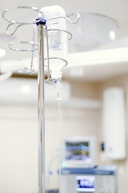
V. Equipment and Supplies for USGIV
Successful USGIV requires specific equipment beyond traditional IV insertion supplies. A portable ultrasound machine with appropriate frequency probes (typically high-frequency for superficial veins) is essential. Linear probes are commonly used for visualizing peripheral veins.
Necessary supplies include standard IV catheters (20-gauge are frequently utilized in training)‚ sterile gloves‚ chlorhexidine for skin preparation‚ and sterile dressings. Vascular phantom models‚ like silicone blocks (Your Design Medical) or tofu models‚ are crucial for initial skill development. A laptop for asynchronous learning and software like Reacts can enhance training‚ facilitating performance recording and feedback.
V.A. Ultrasound Machine Selection
Choosing the right ultrasound machine is paramount for effective USGIV training. Portability is key‚ allowing for bedside application and flexible training environments. Machines should offer high-resolution imaging‚ crucial for visualizing superficial veins. Frequency selection is vital; higher frequencies (7-13 MHz) provide better resolution for smaller vessels‚ while lower frequencies offer deeper penetration.

Consider machines with features like Doppler capability for assessing blood flow and image storage for documentation and review. Cost-effectiveness is also a factor‚ balancing image quality with budgetary constraints. Dedicated vascular ultrasound machines are ideal‚ but general-purpose machines with vascular presets can suffice for training purposes.
V.B. Probe Types and Their Applications
Linear probes are the workhorse for USGIV‚ offering high-frequency‚ high-resolution imaging ideal for superficial veins. Their small footprint facilitates scanning in tight spaces. Curved probes‚ with lower frequencies‚ are less commonly used but can be helpful for larger patients or deeper vessel assessment.
Probe selection impacts image quality and depth of penetration. Higher frequency probes (7-13 MHz) visualize smaller vessels with greater detail‚ crucial for peripheral IVs. Understanding probe characteristics is vital for accurate vein identification. Proper probe care‚ including disinfection and storage‚ is essential to maintain image quality and prevent infection.
V.C. IV Catheters and Accessories
Selecting appropriate IV catheters is crucial for successful USGIV. 20-gauge catheters were utilized in initial training programs‚ demonstrating feasibility with vascular phantom models. Catheter length should match vein depth‚ minimizing the risk of dislodgement. Accessories like sterile gloves‚ chlorhexidine swabs‚ and transparent dressings are essential for maintaining asepsis.
Extension sets facilitate medication administration and reduce catheter trauma. Securement devices prevent accidental dislodgement‚ enhancing patient safety. Understanding catheter materials and flow rates is vital for optimal fluid delivery. Proper disposal of used catheters and accessories adheres to infection control protocols.
VI. Anatomical Considerations for USGIV
Successful USGIV requires a thorough understanding of upper extremity venous anatomy. Key veins include the basilic‚ cephalic‚ and median cubital‚ each presenting unique accessibility and depth considerations. Nurses must identify anatomical landmarks and potential variations‚ recognizing that vascular structures aren’t always consistent.
Precise knowledge is vital to avoid arterial puncture‚ a potentially serious complication. Ultrasound imaging helps differentiate veins from arteries based on compressibility and flow characteristics. Understanding surrounding nerve and tendon locations is also crucial for safe needle placement. Careful scanning and anatomical awareness minimize risks and optimize insertion success.
VI.A. Vein Identification – Basilic‚ Cephalic‚ Median Cubital
Accurate vein identification is paramount for successful USGIV. The basilic vein‚ located medially‚ is often larger but deeper‚ presenting a higher risk of arterial or nerve proximity. The cephalic vein‚ laterally positioned‚ is generally easier to access but may be smaller. The median cubital vein‚ antecubital fossa’s central vein‚ offers a balance of size and accessibility.
Ultrasound distinguishes these veins by their appearance – typically hypoechoic and compressible. Nurses must learn to recognize each vein’s characteristic location and depth. Proper probe positioning and image optimization are essential for clear visualization. Consistent practice with anatomical landmarks builds confidence and proficiency in vein identification.
VI.B. Vascular Anatomy Variations
Human vascular anatomy exhibits significant individual variation. Nurses performing USGIV must anticipate these differences to avoid complications. Vein size‚ depth‚ and branching patterns can deviate substantially from standard anatomical depictions. Some patients may present with aplastic or hypoplastic veins‚ requiring alternative access sites.
Prior medical history‚ including previous IV placements or surgeries‚ can influence vascular anatomy. Thorough ultrasound assessment is crucial to identify these variations before attempting cannulation. Recognizing collateral vessels and atypical vein courses enhances procedural safety and success. Adapting technique based on individual anatomy is a core skill for competent USGIV practice.
VI.C. Avoiding Arterial Puncture
Arterial puncture is a significant complication of IV insertion‚ and USGIV greatly reduces this risk‚ but vigilance is essential. Ultrasound clearly differentiates arteries from veins based on pulsatility; arteries exhibit a waveform‚ while veins are non-pulsatile. Nurses must meticulously assess the vessel’s characteristics before needle insertion.
Proper probe positioning and angle are critical for accurate visualization. Understanding Doppler principles aids in confirming venous flow and identifying arterial signals. Maintaining a shallow insertion angle minimizes the risk of traversing the artery. If arterial puncture occurs‚ immediate pressure application and assessment are paramount. Consistent practice and anatomical knowledge are key to avoidance.
VII. USGIV Technique: Step-by-Step Guide
Successful USGIV requires a systematic approach. First‚ position the patient comfortably and prepare the insertion site with sterile technique. Next‚ perform ultrasound scanning to identify a suitable vein – basilic‚ cephalic‚ or median cubital – assessing its depth and diameter. Visualize the vein in short axis to confirm its structure.
With the probe stabilized‚ advance the IV catheter under real-time ultrasound guidance‚ visualizing the needle tip as it enters the vein. Confirm cannulation by observing a flash of blood in the catheter hub and a clear visualization of the catheter tip within the vessel. Finally‚ remove the needle and secure the catheter‚ verifying patency with saline flush.
VII.A. Patient Positioning and Preparation
Optimal patient positioning is crucial for successful USGIV. The arm should be extended and supported‚ ideally with the wrist slightly hyperextended to enhance vein visibility; Ensure adequate lighting and a comfortable environment to minimize patient anxiety. Thoroughly explain the procedure to the patient‚ addressing any concerns and obtaining informed consent.
Prior to scanning‚ meticulously prepare the insertion site using sterile technique. This includes skin antisepsis with chlorhexidine or povidone-iodine‚ followed by sterile draping. Proper preparation minimizes the risk of infection and ensures a clear ultrasound image. Confirm patient allergies and any contraindications before proceeding.
VII.B. Ultrasound Scanning and Vein Assessment
Begin the scan with the high-frequency linear probe‚ applying gentle pressure to visualize superficial veins. Identify the basilic‚ cephalic‚ or median cubital veins‚ assessing their diameter‚ depth‚ and straightness. Look for vessels that are compressible and pulsatile‚ indicating adequate blood flow. Dynamic assessment‚ involving slight pressure‚ helps differentiate veins from arteries.
Optimize image settings – depth‚ gain‚ and focus – for clear visualization. Scan along the entire length of the chosen vein to identify any tortuosity or valves. Document the vein’s characteristics and location. Proper scanning technique is paramount for accurate vein assessment and successful cannulation.
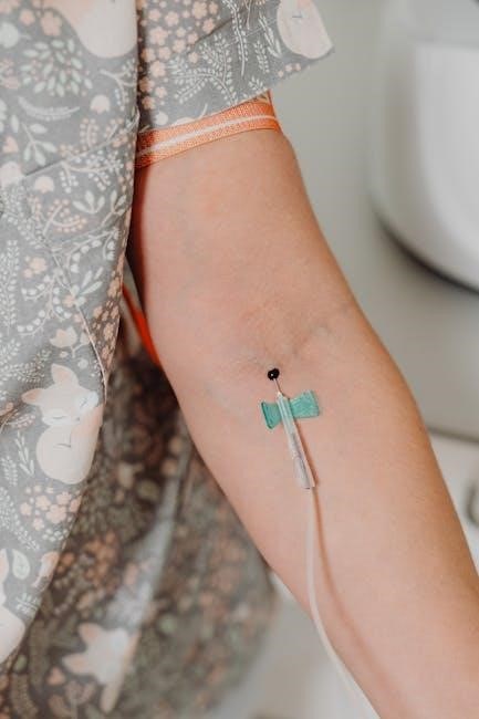
VII.C. Catheter Insertion and Needle Visualization
With continuous ultrasound guidance‚ insert the IV catheter at a shallow angle‚ visualizing the needle tip as it enters the vein lumen. Maintain real-time visualization throughout the insertion process‚ confirming the needle’s trajectory and position within the vessel. Observe for a “flash” of blood into the catheter hub under ultrasound‚ indicating successful cannulation.
Advance the catheter slightly into the vein‚ ensuring the tip is fully within the lumen. Avoid advancing too far‚ which could lead to vessel wall perforation. Throughout the procedure‚ maintain a steady hand and communicate clearly with the patient. Precise needle visualization is crucial for minimizing complications.
VII.D. Confirmation of Cannulation and Removal of Needle
After catheter advancement‚ confirm successful cannulation by observing blood return into the catheter hub under ultrasound guidance. Gently apply pressure proximal to the insertion site while slowly withdrawing the needle. Maintain visualization to ensure the catheter remains within the vein during needle removal.

Secure the catheter according to institutional protocols. Assess for any signs of infiltration or hematoma formation. Document the procedure‚ including the vein accessed‚ number of attempts‚ and any complications encountered. Proper confirmation and securement are vital for maintaining IV access and preventing adverse events.

VIII. Simulation-Based Mastery Learning in USGIV Training
Simulation-based mastery learning is crucial for safe and effective USGIV training‚ requiring participants to demonstrate competency before performing on patients. This approach minimizes risks associated with invasive procedures like peripheral intravenous catheter (PIVC) insertion. Utilizing vascular phantom models – both commercial silicone blocks and cost-effective tofu models – provides realistic practice.
This method‚ supported by studies from Amick et al.‚ emphasizes achieving a minimum passing standard. Performance assessment‚ coupled with constructive feedback‚ is integral. Establishing clear‚ measurable standards ensures nurses possess the necessary skills for successful and complication-free USGIV implementation.
VIII.A. Utilizing Vascular Phantom Models (Silicone & Tofu)
Vascular phantom models are essential tools in USGIV training‚ offering a safe and repeatable environment for skill development. Commercial silicone blocks‚ like those from Your Design Medical‚ provide durable and realistic simulation. However‚ cost-effectiveness is also vital.
Homemade tofu models present a viable alternative‚ offering comparable anatomical features for needle visualization and catheter insertion practice. Both model types allow trainees to repeatedly practice locating veins‚ employing proper ultrasound technique‚ and confirming cannulation without risk to actual patients.
These models facilitate the development of psychomotor skills and build confidence before transitioning to live patient scenarios.
VIII.B. Performance Assessment and Feedback
Robust performance assessment is crucial within simulation-based mastery learning for USGIV. Utilizing tools like Reacts Software allows for objective recording and evaluation of trainees’ technique during simulated IV placements.
Key assessment criteria should include accurate vein identification‚ correct probe positioning‚ successful needle visualization‚ and appropriate catheter insertion angles. Constructive feedback‚ delivered immediately post-simulation‚ is paramount for skill refinement.
This feedback should address both strengths and areas for improvement‚ focusing on technical aspects and adherence to established protocols. Regular assessment and individualized feedback accelerate learning and ensure competency.
VIII.C. Establishing Minimum Passing Standards
Given the potential risks associated with peripheral intravenous catheter (PIVC) insertion‚ implementing minimum passing standards is a logical and essential component of USGIV training. Simulation-based mastery learning necessitates demonstrable competency before progressing to live patient practice.
These standards should be clearly defined and objectively measurable‚ encompassing successful cannulation rates on vascular phantom models – both silicone and tofu – within a specified number of attempts.
Criteria might include consistent visualization of the needle‚ correct catheter placement‚ and absence of simulated complications. Establishing these benchmarks ensures nurses possess the necessary skills to safely and effectively perform USGIV.
IX. Integrating USGIV into Nursing Curriculum
Successful implementation of USGIV requires thoughtful integration into existing nursing educational programs. This isn’t merely an add-on skill‚ but a paradigm shift in IV access‚ demanding dedicated curriculum space.
Initial training should encompass ultrasound physics fundamentals‚ vascular anatomy‚ and hands-on practice with simulation models – silicone and tofu – alongside didactic lectures.
Consider incorporating asynchronous learning modules‚ utilizing pre-recorded videos and performance recording software like Reacts‚ allowing for independent study and self-assessment.
Furthermore‚ ongoing competency assessment and opportunities for skill maintenance are crucial for sustained proficiency in USGIV techniques.
X. Ongoing Competency Assessment and Maintenance
Maintaining proficiency in USGIV necessitates a robust system of ongoing competency assessment. Initial certification isn’t sufficient; regular evaluation ensures skills remain sharp and patient safety is prioritized.
Periodic performance evaluations‚ utilizing vascular phantom models‚ should be implemented to assess scanning technique‚ catheter insertion accuracy‚ and appropriate image interpretation.
Consider incorporating case-based scenarios and simulated clinical challenges to evaluate decision-making skills in complex patient presentations.
Furthermore‚ access to continuing education opportunities and refresher courses is vital for staying abreast of evolving best practices and technological advancements in USGIV.
XI. Troubleshooting Common Challenges in USGIV
Despite training‚ nurses may encounter difficulties during USGIV procedures. Addressing these proactively is crucial for maintaining confidence and minimizing complications.
Common challenges include difficulty visualizing deeper veins‚ particularly in obese patients‚ or those with limited subcutaneous tissue. Recognizing and adjusting probe pressure and frequency are key.
Troubleshooting also involves managing anatomical variations‚ such as tortuous vessels or aberrant branching patterns.
Furthermore‚ recognizing and mitigating artifacts – echoes that don’t represent true anatomy – is essential for accurate interpretation.
Dedicated training modules focusing on these specific challenges‚ coupled with mentorship opportunities‚ will empower nurses to overcome obstacles effectively.
XII. Legal and Ethical Considerations of USGIV Practice
Implementing USGIV requires careful consideration of legal and ethical implications. Nurses must operate within their scope of practice‚ adhering to institutional policies and state regulations regarding advanced skills.
Informed consent is paramount; patients must understand the benefits and risks of USGIV compared to traditional methods. Documentation must be meticulous‚ detailing the indication‚ vein selection‚ procedure details‚ and any complications encountered.

Competency assessment is ethically vital – nurses should only perform USGIV after demonstrating proficiency through validated training and ongoing evaluation.
Liability concerns necessitate adherence to established protocols and a commitment to continuous quality improvement. Maintaining patient confidentiality and advocating for their best interests remain central ethical obligations.
XIII. Future Trends in USGIV for Nursing
The future of USGIV in nursing points towards increased integration of technology and expanded accessibility. Expect advancements in portable‚ cost-effective ultrasound devices‚ making USGIV more readily available in diverse clinical settings.
Artificial intelligence (AI) may assist with vein identification and guidance‚ enhancing accuracy and efficiency. Tele-ultrasound‚ where experienced practitioners remotely guide less experienced nurses‚ could bridge skill gaps.
Curriculum development will likely emphasize augmented reality (AR) and virtual reality (VR) simulations for enhanced training.

Further research will focus on optimizing catheter selection and insertion techniques‚ minimizing complications‚ and demonstrating long-term cost-effectiveness. Wider adoption hinges on establishing clear national standards and advocating for appropriate reimbursement models.